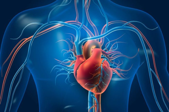
The heart is a solid organ with the ability to coordinate the circulatory design. It is set in the mark of assembly of the chest, in the front mediastinum and, covered by the pericardium, is separated into four pits, the two atria, liable for getting blood from the venous dispersal and the two ventricles, which can siphon blood into the courses.
Anatomy Of The Heart, A Journey Around Life
The legitimate ventricle liberates deoxygenated blood through the tricuspid valve from the right chamber into the respiratory vein. Shockingly, the left ventricle dispatches oxygenated blood from the aspiratory dispersal into the aorta, secured from the left region through the mitral valve.
The interventricular septum separates the two ventricles. The fitting room gets deoxygenated blood from the general course using the certain vena cava, not excellent vena cava, and coronary ostium; the left section helps oxygenated blood through the pneumonic veins. Like this, venous blood is poor in oxygen courses in the sensible bits of the heart; the opposite technique for getting around in the left district, the blood is a vein and oxygenated.
Wanting to work on cardiovascular headway in whatever amount as could sensibly be anticipated, we can say that the oxygen-awful blood of the general stream shows up at the adequate chamber from where, through the tricuspid valve, it passes to the right ventricle, which siphons it, through the pneumonic aide, into the lungs, where is oxygenated. From the lungs, the blood appears, through the respiratory veins, in the left chamber, where the mitral valve passes into the left ventricle, which releases it into the aorta passage. The ventricles are associated with the veins utilizing the aortic valve on the left and the inspiratory valve on the right.
In the circulatory system, there are four helpers in a social gathering; the two atria, whose limit is to siphon blood into the ventricles, and the two ventricles, with a more significant siphon limit. The valves grant the dissemination framework to occur in a lone bearing, hindering reflux. The left ventricle has essentially higher muscle wall thickness than the contralateral one, in this way having the choice to convey higher strains and introducing considerations of electrocardiography, making conceivable electrical aftereffects of more fundamental plentifulness.
Cardiac Ultrasound And Electrocardiogram
The contractile action of cardiovascular muscle cells, the siphon ability of which we have, as of late analyzed, happens thanks to the presence of the electrical conduction structure, which in clinical practice is focused on through the electrocardiogram. The mechanical development of the heart, the valve ability, the vessels, and the pericardium can be consistently inspected by ultrasound.
The electrical conduction system is formed by unambiguous cells, with the piece of a pacemaker and the spread of the electrical redesign. Physiologically, the electrical inspiration begins at the sinus center level, the fundamental heart pacemaker, arranged in the right chamber; from here, it spreads concentrically, depolarizing the atria and allowing them to get; this characteristic is recorded electrocardiographically as a P wave.
Through the internodal conduction pathways, the electrical inspiration shows up at the atrioventricular center, which is dealt with through a quick pause, essential for blood from the atria to the ventricles. As we will find in the future while examining dull and brady arrhythmias, the ability of the atrioventricular center anticipates pivotal importance. This brief pause connects with the ECG following the Pr part. A short time later, the electrical drive journeys extraordinarily rapidly along a load of His and the right and left branches, depolarizing the ventricles and allowing them to get; this connects with the QRS complex of ECG following. Finally, there is repolarization of the ventricles, which, after the St part, looks at the T wave.
This time of heart idleness is fundamental in clinical practice since an ectopic electrical drive, for instance, made outer the Sa center point, which goes to electrically empowering the myocardium in this stage, would have a basic jeopardize of causing extremely dangerous ventricular arrhythmias. To accurately grasp cardiovascular movement, physiological or obsessive, it is fundamental to consider the mechanical activity of the heart stringently synergistic with the electrical one.
To move toward arrhythmias, which we will examine in later articles, it is essential to comprehend that every heart cell can accept the job of a pacemaker and can direct the electrical upgrade; if this were not the situation, any modification of the conduction tissue, continuous in clinical practice, would have extreme repercussions on the working of the heart.
Legitimately, the pacemaker capability of the cells at first not relegated to this action will have substantially less viability for the sinus hub the further away from the atria towards the ventricles; this will show itself with a much lower beat release recurrence passing from the atrial cells, to the Av hub, to the cells of the heap of His, to the ventricular myocardial cells; generally, the hub Sa releases beat at a recurrence of 60-100/m’; the Nav 45-50; myocardial cells 30-35/m’. Simultaneously, the engendering of the electrical drive, which is exceptionally quick through the common pathways, will likewise be dialed back on account of pathologies influencing the conduction pathways.
Also Read: Physical Activity After A Heart Attack

